
|
Upload | Sign in |
|---|
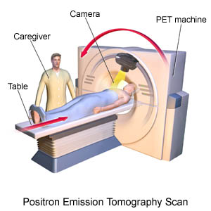
PET (Positron Emission Tomography)
A PET scan uses radiation, or nuclear medicine imaging, to produce 3-dimensional, colour images of the functional processes within the human body.
The PET principle is based on the phenomenon of positron annihilation that gives rise to gamma rays i.e two photons are simultaneously emitted in almost exactly opposite directions whenever a positron passes through matter.
The use of positron emitters for medical imaging purposes was first suggested by Wrenn et al. and Sweet in the early 1950s.
Recently, the Positron Emission Tomography (PET) system, coupled with a CT scan, is performed on the patient during the same session, using the same machine.
The CT images are matched with the PET scans. This technique improves the anatomic precision of the displayed images and provides a means for the nuclear count correction.
PET scans are increasingly read alongside CT or magnetic resonance imaging (MRI) scans, the combination giving both anatomic and metabolic information.
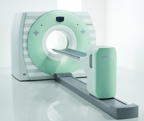
Positron Emission Tomography (PET) imaging is most useful in combination with anatomical imaging, such as CT, modern PET scanners are now available with an integrated CT scanner. Because the two scans can be performed in immediate sequence during the same session, with the patient not changing position between the two types of scans, the two sets of images are more precisely registered, so that areas of abnormality on the PET imaging can be more perfectly correlated with anatomy on the CT images.
This is very useful in showing detailed views of moving organs or structures with higher anatomical variation.
GENERATION’s:
The first positron computed tomography machine was developed in 1975 by Ter-Pogossian et al.
This system was referred to as positron emission transaxial tomography (PETT II), which consisted of a hexagonal array of NaI (T1) detectors connected in coincidence between opposite pairs.
The filtered back projection (FBP) reconstruction method was adopted in that system.
The first whole body positron computed tomography (PETT III) was developed shortly thereafter and it was used in human studies.
Principle:
.JPG)
The radioactive decay mechanism for positron emitters used in PET is positron emission, whereby a proton in the nucleus is transformed into a neutron and a positron.
The positron (β+) has exactly the same mass and same magnitude of charge as the electron except that the charge being carried is positive.
For positron emission to be energetically feasible, the total energy difference between the parent and the daughter states should be at least 1.022 MeV. Positron emitters are of special interest in medicine because the main elements (e.g. carbon, oxygen and nitrogen) that constitute living organisms have isotopes that emit positrons.
The positron will have some initial energy after emission from the parent nucleus.
It travels a short distance from the nucleus, scatters and collides with loosely bound electrons nearby before fusing with one of them to form positronium and then annihilates.
Their mass converts into energy in the form of two 511 keV emitted 180° to each other of these two 511 keV photons forms the basis of PET imaging. The probability that both 511 keV photons will escape from the body without scattering is very high and both photons can be detected with two detectors at opposite ends of the line.
.JPG)
APPLICATIONS:
PET scans are commonly used to investigate the following conditions:
Epilepsy
Alzheimer's disease
Cancer
Heart disease
Medical research
blog comments powered by Disqus